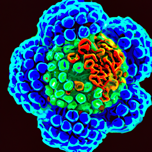
Have you ever wondered how scientists are able to visualize viruses, those tiny, invisible entities that can cause so much havoc? Well, the answer lies in the remarkable technology of optical microscopes. With their ability to magnify objects up to a thousand times, these powerful tools play a vital role in helping scientists study and understand the intricate structures and behaviors of viruses. In this article, we will explore the fascinating world of optical microscopy and uncover the secrets behind its contribution to the field of virology. So get ready to journey into the microscopic realm and unlock a whole new perspective on the tiny organisms that shape our world.
Introduction
Welcome to this comprehensive article on optical microscopes and their role in visualizing viruses. In this article, we will explore the basics of optical microscopes, their limitations, various techniques for visualizing viruses, case studies of virus visualization, recent advances in optical microscopy, and the future perspectives of this field. By the end of this article, you will have a deeper understanding of how optical microscopes contribute to virus detection and diagnosis, as well as their broader applications in biomedical research.
I. Basics of Optical Microscopes
A. Definition and Function
Optical microscopes, also known as light microscopes, are essential tools in the field of microscopy. Their primary function is to visualize and magnify objects that are not visible to the naked eye. Unlike electron microscopes that use electrons to create an image, optical microscopes use visible light to illuminate the sample and produce an enlarged image. These microscopes are widely used in various scientific disciplines, including biology, medicine, materials science, and forensic science.
B. Components of an Optical Microscope
An optical microscope consists of several key components that work together to produce a magnified image. These components include an objective lens, an eyepiece lens, an illuminator, a condenser lens, and a stage. The objective lens is responsible for capturing the light from the sample and producing the initial image. The eyepiece lens further magnifies the image, allowing the viewer to see the details more clearly. The illuminator provides the light source, and the condenser lens focuses the light onto the sample. The stage holds the sample in place and allows for precise movement and positioning.
C. Working Principle of Optical Microscopes
The working principle of an optical microscope involves the interaction of light with the sample. When light passes through the objective lens, it interacts with the sample, causing some of the light to be absorbed, reflected, or scattered. The remaining light passes through the eyepiece lens, where it is further magnified and focused to create an enlarged image. The image is then observed by the viewer through the eyepiece. By adjusting the focus, illumination, and other parameters, optical microscopes can provide a clear and detailed visualization of the sample.

II. Limitations of Optical Microscopes
A. Size Limitations
One of the limitations of optical microscopes is their inability to visualize objects that are smaller than the wavelength of visible light. This is due to the diffraction limit, which determines the smallest resolvable detail of an optical microscope. As viruses are typically smaller than the diffraction limit, it is challenging to directly visualize them using conventional optical microscopy techniques. However, there are alternative techniques that can overcome this limitation, which we will discuss later in this article.
B. Resolution Limitations
Another limitation of optical microscopes is their limited resolution, which refers to the ability to distinguish between two closely spaced objects. The resolving power of an optical microscope is determined by the numerical aperture of the objective lens and the wavelength of the illumination source. High-resolution imaging of viruses requires techniques that can surpass the diffraction limit of conventional optical microscopy, such as super-resolution microscopy.
C. Contrast Limitations
Optical microscopes rely on the contrast between the sample and its surroundings to produce clear images. However, viruses often have low inherent contrast, making them difficult to distinguish from the background. Staining techniques, discussed in the next section, can enhance the contrast by selectively labeling the viruses or their components, enabling better visualization.
III. Techniques for Visualizing Viruses
A. Staining
Staining is a common technique used in optical microscopy to enhance the contrast of the sample. It involves selectively applying dyes or fluorescent molecules that bind to specific components of the viruses. By choosing the appropriate stain, researchers can highlight the viruses and distinguish them from the background. Staining techniques have been widely used to visualize various viruses, including HIV, influenza, and herpes, as we will explore in the following section.
B. Fluorescence Microscopy
Fluorescence microscopy is a powerful technique that utilizes fluorescent molecules to visualize viruses. By labeling the viruses with fluorescent probes, researchers can observe their location, distribution, and interaction with other cellular components. Fluorescence microscopy offers high specificity and sensitivity, enabling the detection and tracking of viruses in complex biological samples. This technique has significantly contributed to our understanding of viral infection mechanisms and host-virus interactions.
C. Labeling
Labeling refers to the process of attaching a marker to the viruses or their components to facilitate their detection and visualization. This can be achieved by using specific antibodies that bind to viral proteins or by genetically modifying the viruses to express fluorescent proteins. The labeled viruses can then be visualized using optical microscopes, allowing researchers to study their behavior and characteristics.
D. Immunohistochemistry
Immunohistochemistry is a widely used technique that combines immunological methods with optical microscopy. By utilizing specific antibodies that bind to viral antigens, researchers can identify and localize viruses within tissue samples. Immunohistochemistry provides valuable information about the distribution and abundance of viruses in various organs and tissues, aiding in the diagnosis and understanding of viral diseases.

IV. Case Studies of Virus Visualization
A. HIV
Visualization of the Human Immunodeficiency Virus (HIV) has played a crucial role in advancing our understanding of this devastating disease. Through the use of fluorescent labeling and immunohistochemistry techniques, researchers have been able to track the progression of HIV infection, study the viral replication cycle, and identify potential targets for antiviral therapies. Furthermore, optical microscopy has been instrumental in the development of diagnostic tests for detecting HIV in patient samples.
B. Influenza
Influenza viruses, responsible for seasonal flu outbreaks, have been extensively visualized using optical microscopy techniques. Staining and fluorescence microscopy have helped researchers study the structure, replication, and transmission of influenza viruses. By visualizing the interaction of influenza viruses with host cells, scientists have gained insights into the pathology of the disease and developed strategies for vaccine development and antiviral drug discovery.
C. Herpes
Herpes viruses, including herpes simplex virus (HSV), have been studied using a combination of staining and immunohistochemistry techniques. Optical microscopy has allowed researchers to visualize the viral particles, track their entry into host cells, and study the establishment of latency. These studies have contributed to our understanding of herpes infection mechanisms and have implications for the development of antiviral therapies.
V. Recent Advances in Optical Microscopy
A. Super-resolution Microscopy
Super-resolution microscopy is an emerging technique that overcomes the diffraction limit of conventional optical microscopes. By utilizing advanced imaging principles and fluorescent probes, super-resolution microscopy can achieve resolutions below the diffraction limit, enabling the visualization of viruses and their subcellular components with unprecedented detail. This technique has revolutionized the field of virology and has opened up new avenues for studying viral structures and dynamics.
B. Electron Microscopy Combined with Light Microscopy
Combining the benefits of both electron microscopy and light microscopy has led to significant advancements in virus visualization. By using electron microscopy to observe viruses at ultra-high resolutions, researchers can obtain detailed structural information. Additionally, fluorescence labeling techniques can be employed to localize specific viral components, providing valuable insights into their function and interactions within the infected host cells.
C. Bessel Beam Illumination
Bessel beam illumination is a novel technique that allows for the imaging of viruses in three dimensions without the need for scanning. Unlike traditional microscopy techniques that use point scanning, Bessel beam illumination generates a non-diffracting beam that can illuminate the entire sample simultaneously. This three-dimensional imaging capability has the potential to reveal the dynamic behavior of viruses within living cells, enhancing our understanding of viral replication and pathogenesis.
D. Light Sheet Microscopy
Light sheet microscopy, also known as selective plane illumination microscopy (SPIM), has emerged as a powerful technique for imaging viruses in living specimens. By illuminating the sample with a thin sheet of light, light sheet microscopy reduces phototoxicity and provides high-resolution imaging. This technique has been successfully applied to visualize virus infection dynamics in real-time, enabling researchers to study the temporal and spatial aspects of viral replication within living organisms.
VI. Future Perspectives
A. Development of New Techniques
The field of optical microscopy continues to evolve, with ongoing research focused on developing new techniques and improving existing ones. Scientists are exploring novel imaging modalities, such as single-molecule imaging and correlative light-electron microscopy, to further enhance the visualization of viruses. These advancements will provide researchers with even higher resolution and more detailed information about viral structures and dynamics.
B. Integration with Other Technologies
The integration of optical microscopy with other technologies, such as artificial intelligence and machine learning, holds great promise for the field of virus visualization. By combining image analysis algorithms with the vast amount of data generated by optical microscopes, researchers can automate virus detection, classification, and quantification. This integration will significantly speed up the diagnostic process and enhance our ability to study viral infections on a large scale.
C. Application in Biomedical Research
Optical microscopy techniques are not only limited to virus visualization but also find extensive applications in biomedical research. By visualizing viruses in the context of host cells and tissues, researchers can gain insights into the mechanisms of viral pathogenesis, host immune responses, and potential therapeutic targets. Optical microscopy will continue to be a valuable tool in advancing our understanding of viral diseases and developing effective treatment strategies.
D. Potential for Virus Detection and Diagnosis
As optical microscopy techniques continue to improve, there is great potential for their application in virus detection and diagnosis. By harnessing the advancements in staining, labeling, and super-resolution imaging, optical microscopes can become powerful diagnostic tools in clinical settings. Rapid and accurate identification of viral infections can aid in early intervention, treatment selection, and outbreak control. Furthermore, optical microscopy techniques can be integrated into point-of-care devices for on-site virus detection, especially in resource-limited settings.
VII. Conclusion
In conclusion, optical microscopes have revolutionized the field of virus visualization, enabling researchers to study viruses at various levels of detail. Despite their limitations, optical microscopes, coupled with staining, fluorescence, and immunohistochemistry techniques, have played a critical role in advancing our understanding of viral infections. Recent advancements in optical microscopy, such as super-resolution microscopy, electron microscopy combined with light microscopy, and novel illumination techniques, have further expanded our capabilities for virus visualization. With continued development, integration with other technologies, and applications in biomedical research and diagnostics, optical microscopy will continue to be an invaluable tool in combating viral diseases and improving human health.


