In the world of microscopy, two powerful tools have revolutionized our understanding of the microscopic world: the optical microscope and the electron microscope. While both devices allow us to glimpse into the realm of minuscule structures and organisms, they employ vastly different principles and technologies. In this article, we will explore the differences between these two types of microscopes and discuss the unique advantages they offer in exploring the hidden wonders of the microscopic world. Whether you’re a science enthusiast, a student, or simply curious about the extraordinary world that exists beyond our naked eye, this comparison will shed light on the fascinating world of microscopy.
Comparing Optical Microscope and Electron Microscope
1. Introduction
1.1 Purpose of the article
The purpose of this article is to provide a comprehensive overview and comparison of the optical microscope and the electron microscope. Both of these microscopes are incredibly useful tools in the field of microscopy, but they differ in their principles, imaging techniques, sample preparation, magnification, observational capabilities, cost, and accessibility. By understanding the characteristics and differences of these two types of microscopes, you can make an informed decision about which one is better suited for your specific needs.

2. Overview of Optical Microscope
2.1 Definition
An optical microscope, also known as a light microscope, is an instrument that uses visible light and a series of lenses to magnify small objects and generate a magnified image. It is one of the oldest and most widely used types of microscopes.
2.2 Basic Principles
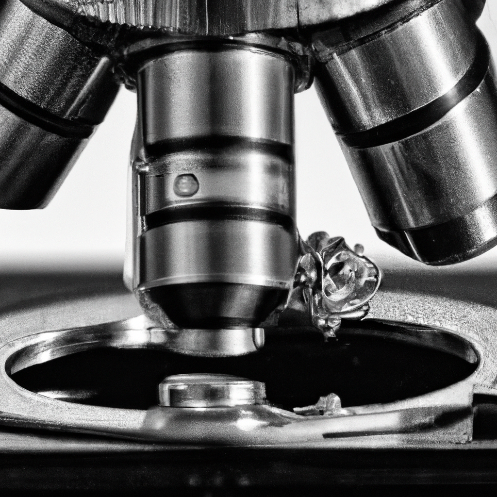
The basic principle of an optical microscope involves passing visible light through the sample and using a series of lenses to magnify and focus the light, allowing the human eye or a camera to observe the sample. The lenses in an optical microscope include the objective lens, which is located close to the sample, and the eyepiece lens, which is positioned near the observer’s eye. The magnification of an optical microscope is determined by the combination of these lenses.
2.3 Components
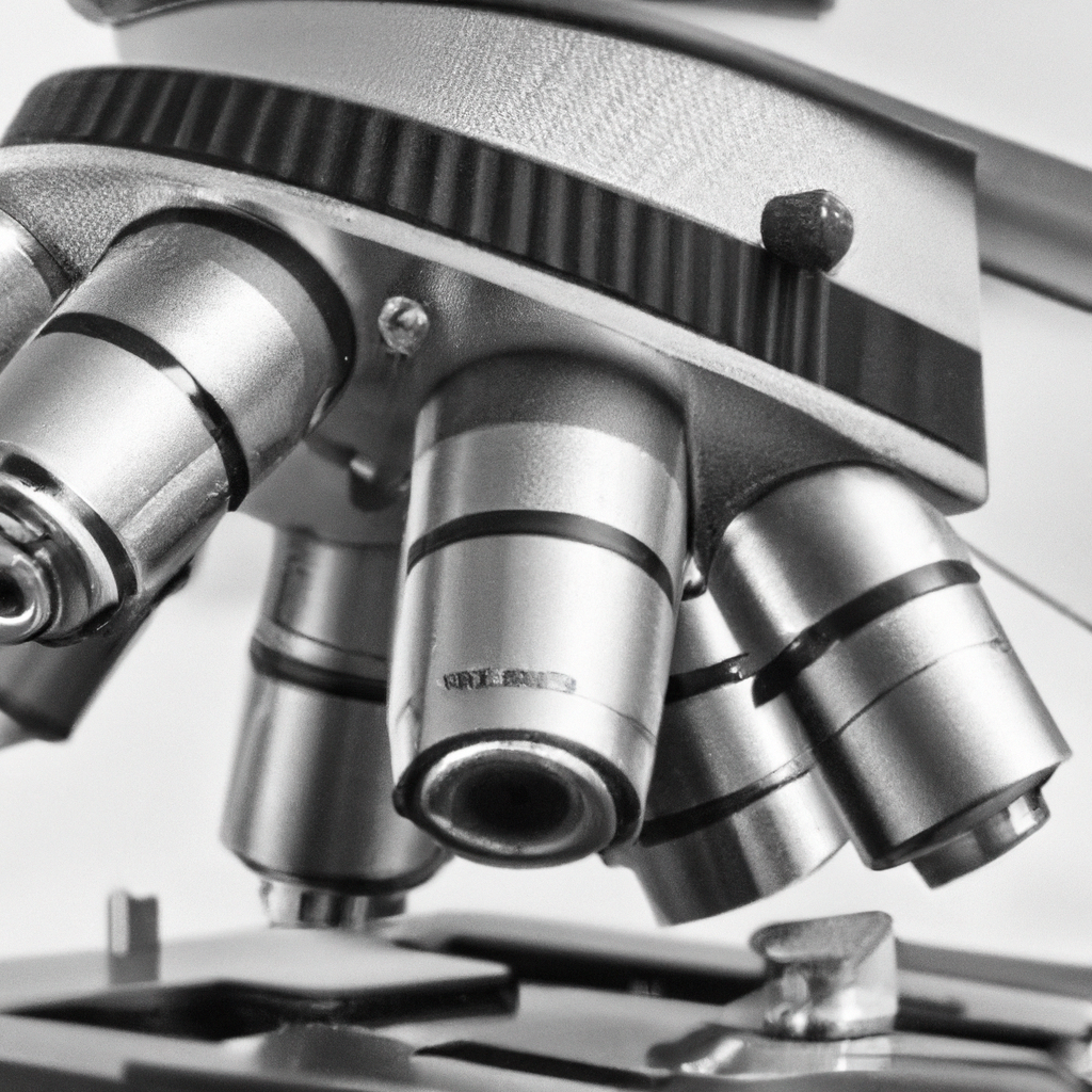
The components of an optical microscope typically include an illumination system to provide light, a stage to hold the sample, a set of objective lenses with different magnifications, an eyepiece or a camera for observation or image capture, and various adjustments for focusing the sample.
3. Overview of Electron Microscope
3.1 Definition
An electron microscope is an instrument that uses a beam of electrons instead of visible light to illuminate the sample and create highly detailed and magnified images. It is a more recent invention compared to the optical microscope and has revolutionized the field of microscopy.
3.2 Basic Principles
The basic principle of an electron microscope involves accelerating a beam of electrons towards the sample and using electromagnetic lenses to focus the electrons onto the sample. The interaction of the electrons with the sample produces signals that are detected and used to generate an image. The electrons used in electron microscopes have much shorter wavelengths than visible light, allowing for much higher resolution imaging.
3.3 Types of Electron Microscopes
There are different types of electron microscopes, including the transmission electron microscope (TEM) and the scanning electron microscope (SEM). The TEM allows for imaging the internal structure of a sample by transmitting electrons through it, while the SEM scans the sample’s surface with a focused electron beam, providing information about the sample’s topography and composition.

4. Differences in Imaging
4.1 Imaging Technique
The imaging technique differs significantly between optical microscopes and electron microscopes. Optical microscopes use visible light to illuminate the sample, and the image is observed directly by the eye or a camera. In contrast, electron microscopes use a beam of electrons to scan or pass through the sample, and the resulting signals are detected and used to generate an image on a screen.
4.2 Resolution
One of the key differences between optical microscopes and electron microscopes is resolution. Optical microscopes have a limited resolution due to the wavelength of visible light, typically around 200-300 nanometers. On the other hand, electron microscopes have much higher resolution capabilities, reaching sub-nanometer scales. This enables the visualization of ultrafine details in a sample not visible with optical microscopes.
4.3 Depth of Focus
The depth of focus, or depth of field, refers to the range of distances from the microscope lens within which the sample remains in focus. Optical microscopes generally have a greater depth of focus compared to electron microscopes, allowing for a larger portion of the sample to be in focus simultaneously. However, electron microscopes can achieve a high level of precision in focusing on specific areas of interest within the sample.
5. Differences in Sample Preparation
5.1 Sample Types
Sample preparation methods vary depending on the type of microscope used. Optical microscopes can handle a wide range of samples, including biological specimens, thin films, and solid materials. Electron microscopes, on the other hand, require more meticulous sample preparation. Samples often need to be dehydrated, fixed, embedded in resin, and sliced into thin sections for transmission electron microscopy. Surface coating with conductive materials may also be necessary for scanning electron microscopy.
5.2 Sample Preparation Techniques
Optical microscopy typically requires minimal sample preparation, with simple techniques such as mounting the sample on a glass slide and applying a cover slip. However, electron microscopy requires more complex techniques, including chemical fixation, staining, and the use of heavy metal contrasting agents. These preparation techniques are necessary to enhance the sample’s contrast and create a more detailed image.
6. Differences in Magnification and Observational Capabilities
6.1 Magnification Range
When it comes to magnification, electron microscopes have a significant advantage over optical microscopes. Electron microscopes can achieve much higher magnification levels, often exceeding one million times. Optical microscopes, on the other hand, typically have a maximum magnification range of around 1000 times. The high magnification capabilities of electron microscopes allow for the visualization of ultrafine details and structures within the sample.
6.2 Observational Capabilities
The observational capabilities of optical microscopes and electron microscopes also differ. Optical microscopes provide real-time imaging, allowing for the direct observation of dynamic processes in the sample. In contrast, electron microscopes often require more time for sample preparation and imaging, and the process may involve high vacuum conditions. Electron microscopes also offer advanced techniques such as elemental analysis through energy-dispersive X-ray spectroscopy and three-dimensional imaging through electron tomography.
7. Differences in Cost and Accessibility
7.1 Cost
Cost is an important consideration when choosing a microscope. Optical microscopes are generally more affordable and accessible compared to electron microscopes. The lenses and components of optical microscopes are widely available and come at relatively lower costs. On the other hand, electron microscopes are complex and expensive instruments, often requiring specialized facilities and maintenance. The purchase, installation, and upkeep of electron microscopes can be prohibitively expensive for many researchers and institutions.
7.2 Accessibility
Optical microscopes are widely accessible and commonly found in educational institutions, research laboratories, and clinical settings. They are relatively easy to operate, and users do not require specialized training to use them effectively. Electron microscopes, on the other hand, are less accessible due to their high cost, technical complexity, and specialized requirements. They are typically found in specialized research facilities and institutions with dedicated experts and technicians to operate and maintain them.
8. Advantages and Disadvantages of Optical Microscopy
8.1 Advantages
Optical microscopy has numerous advantages. First and foremost, it is generally more affordable and accessible compared to electron microscopy. It is also well-suited for real-time imaging, allowing for the observation of dynamic processes in the sample. Optical microscopes are versatile, capable of analyzing a wide range of samples, including live cells, tissues, and solid materials. They also have a larger depth of focus, making them useful for observing three-dimensional structures.
8.2 Disadvantages
Despite its advantages, optical microscopy does have some limitations. Its resolution is limited by the wavelength of visible light, preventing the visualization of ultrafine details. Optical microscopes also have a lower magnification range compared to electron microscopes, which may limit the level of detail observed. Additionally, optical microscopes may require staining techniques to enhance contrast in certain samples, which can potentially alter the sample’s characteristics.
12. Conclusion
In conclusion, the choice between an optical microscope and an electron microscope depends on your specific requirements and applications. Optical microscopes are affordable, accessible, and suitable for observing a wide range of samples in real-time. They have a larger depth of focus but are limited in resolution and magnification. Electron microscopes, on the other hand, offer superior resolution and magnification capabilities, but they come at a significantly higher cost and require more complex sample preparation. They are particularly useful for studying ultrafine details and conducting advanced analytical techniques. Consider the specific needs of your research or application to make an informed decision between these two powerful microscopy tools.


