Imagine being able to explore the tiny world that exists beyond the naked eye, right from the comfort of your own home or laboratory. With a digital microscope, you can do just that. In this article, we will uncover the fundamentals of a digital microscope, shedding light on its features, applications, and how it works. Get ready to embark on a fascinating journey, where the unseen becomes visible and the microscopic becomes extraordinary.

What is a Digital Microscope
A digital microscope is a modern and advanced tool used for viewing and examining objects that are too small to be seen with the naked eye. It combines traditional microscope technology with digital imaging capabilities, allowing users to capture, store, and share high-resolution images and videos of their specimens. It provides a convenient and efficient way to observe and analyze microscopic details, making it an invaluable tool in various fields such as science, research, medicine, manufacturing, and education.
Working Principle of a Digital Microscope
Optical System
The optical system is the core component of a digital microscope. It consists of an objective lens, an eyepiece, and several mirrors or prisms. The objective lens collects and focuses the light from the specimen, forming a magnified real image. The eyepiece further magnifies this image and allows the user to observe it. The mirrors or prisms in the optical system direct the light path and ensure optimal image quality.
Camera
At the heart of the digital microscope is the camera. It captures the magnified real image produced by the optical system and converts it into digital signals. These signals are then processed and transformed into a digital image that can be displayed on a screen or saved for later analysis. The camera plays a vital role in the overall image quality and resolution of the digital microscope.
Light Source
The light source illuminates the specimen, allowing it to be seen and examined under the microscope. Traditional microscopes typically use halogen or LED lights, but digital microscopes often incorporate advanced LED lighting systems. These systems provide consistent and adjustable illumination, ensuring optimal image clarity and contrast. The light source can be controlled and manipulated to enhance specific details or highlight certain features of the specimen.
Image Processing
Image processing is an essential feature of digital microscopes. It involves the use of specialized software to enhance and refine the captured digital images. This software can adjust brightness and contrast, remove noise and artifacts, and even apply various filters or image enhancement techniques. Image processing helps to improve the overall image quality, making it easier for the user to observe and analyze the microscopic details.
Types of Digital Microscopes
There are several different types of digital microscopes available, each suitable for specific applications and user requirements. These include:
Compound Microscope
The compound microscope is the most common type of microscope and is widely used in scientific research, education, and medical diagnostics. It utilizes a combination of multiple lenses to provide high magnification levels and excellent resolution. Compound microscopes are typically used for observing thin, transparent specimens such as cells, bacteria, and tissue samples.
Stereo Microscope
The stereo microscope, also known as a dissecting microscope, is commonly used for studying larger, solid objects that do not require high magnification. It provides a three-dimensional view of the specimen, allowing for better depth perception and spatial awareness. Stereo microscopes are often employed in fields such as biology, botany, and quality control.
USB Microscope
As the name suggests, a USB microscope connects directly to a computer or other digital device via a USB port. It is a compact and portable option that is ideal for users who need to capture and analyze images on the go. USB microscopes are commonly used in fields like electronics inspection, jewelry appraisal, and hobbyist microscopy.
Digital Handheld Microscope
A digital handheld microscope is a portable and lightweight option that can be held directly over the object of interest. It is equipped with its own built-in display or can connect to a computer or mobile device for image viewing. This type of microscope is popular in fields such as entomology, archaeology, and outdoor exploration.
Digital Inspection Microscope
Digital inspection microscopes are primarily used for quality control and inspection purposes in manufacturing industries. They provide high magnification, precise measurements, and advanced imaging capabilities. These microscopes are often equipped with features such as auto-focus, measurement tools, and image comparison functions.
Key Components of a Digital Microscope
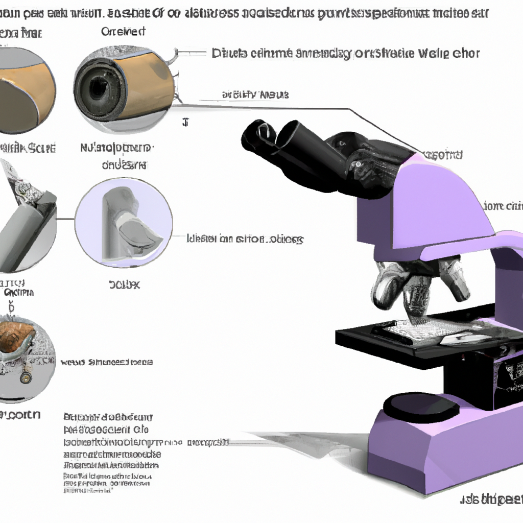
To understand how a digital microscope works, it is essential to familiarize yourself with its key components. These components include:
Objective Lens
The objective lens is the primary lens responsible for magnifying the specimen and collecting light from it. It determines the overall magnification power and spatial resolution of the microscope. Objective lenses are available in various magnification levels and numerical apertures, allowing users to select the appropriate lens for their specific needs.
Eyepiece
The eyepiece, also known as the ocular lens, is located at the top of the microscope and is where the user directly observes the magnified image. It further magnifies the image produced by the objective lens, allowing for detailed observation. Eyepieces usually have a fixed magnification, although some digital microscopes may have adjustable or interchangeable eyepieces.
Digital Camera
The digital camera is the crucial component that captures the magnified image from the objective lens. It may be an integrated camera within the microscope or a detachable camera that can be connected via USB or other interfaces. The camera converts the optical image into digital signals, allowing for real-time viewing and image capture on a screen or computer.
LED or Halogen Light Source
The light source provides illumination to the specimen, allowing it to be visible under the microscope. Digital microscopes often utilize LED lights due to their energy efficiency, adjustable intensity, and long lifespan. However, some high-end microscopes still use traditional halogen lights, which provide a broader spectrum of light for enhanced color representation.
Focusing Mechanism
The focusing mechanism allows the user to adjust the focus of the microscope and bring the specimen into sharp clarity. It may be manual, with knobs or wheels for coarse and fine adjustments, or motorized with automated focusing capabilities. A smooth and precise focusing mechanism is essential for obtaining clear and detailed images.
Stage
The stage is the platform where the specimen is placed for observation. It is usually a flat surface with clips, slides, or other holders to secure the specimen in place. The stage may also have mechanical movements, allowing for precise positioning and scanning of the specimen. Some advanced digital microscopes have motorized or automated stage movements.
Software
The software is a vital component of a digital microscope as it enables advanced image processing, analysis, and control functionalities. It provides features such as image capture, measurement tools, annotation, and sharing options. The software may also allow for image stitching, 3D reconstruction, and time-lapse imaging, depending on the microscope model and capabilities.

Resolution and Magnification
Resolution and magnification are key specifications to consider when evaluating the performance of a digital microscope.
Resolution refers to the level of detail that can be observed and captured by the microscope. It is determined by the number of pixels in the camera sensor and the optical quality of the microscope system. Higher resolution results in sharper and more detailed images, allowing for accurate analysis and observation of microscopic structures.
Magnification, on the other hand, refers to the degree of enlargement of the specimen compared to its actual size. It is determined by the combination of objective lens and eyepiece magnifications. Digital microscopes typically offer a range of magnification options, allowing users to choose the appropriate level of enlargement for their specific needs.
It is important to note that higher magnification does not always result in better image quality. The resolution of the microscope system must be sufficient to support the selected magnification level. Additionally, factors such as lighting, focus, and specimen preparation also play a significant role in the overall image quality and clarity.
Advantages of Digital Microscopes
Digital microscopes offer several advantages over traditional optical microscopes. These advantages include:
Capture and Store Images
One of the most significant advantages of digital microscopes is the ability to capture high-resolution images of specimens. These images can be saved digitally, allowing for easy storage, organization, and retrieval. Digital image files can also be easily shared with colleagues, allowing for collaboration and remote analysis.
Record Videos
Digital microscopes often have the capability to record videos of the observed specimens. This is particularly useful for capturing dynamic processes or movements that cannot be adequately conveyed through static images alone. Video recordings can be reviewed, analyzed, and shared, providing a valuable tool for research, education, and documentation.
Easy Image Sharing
Digital microscopes allow for seamless image sharing. Images captured by the microscope can be instantly shared via email, messaging apps, or online platforms. This facilitates collaboration and remote discussions, enabling researchers, educators, and professionals to easily exchange ideas and observations.
Digital Measurements
Digital microscopes often come with built-in measurement tools or software capabilities that enable accurate measurement of specimens. These measurements can be performed directly on the digital images, eliminating the need for manual measurements using rulers or calipers. This simplifies the analysis process and improves measurement accuracy.
Real-Time Viewing
Digital microscopes provide real-time viewing of the specimen on a screen or monitor. This allows multiple users to observe and discuss the specimen simultaneously, making it ideal for group discussions, teaching, or presentations. Real-time viewing also enables immediate adjustments to the microscope settings for optimal image quality and clarity.
Image Enhancement
Digital microscopes often come with software that allows for image enhancement and manipulation. This includes adjusting brightness, contrast, and color settings, as well as applying filters or image enhancement algorithms. Image enhancement tools help to improve the visibility of specific details or highlight certain features in the image, enhancing the overall observation experience.
Applications of Digital Microscopes
Digital microscopes have a wide range of applications across various fields and industries. Some of the key applications include:
Biological Research
In biological research, digital microscopes are extensively used for studying cell structures, tissues, and organisms. They enable researchers to observe and analyze cellular processes, investigate diseases, and advance our understanding of the living world. Digital microscopes are used in areas such as genetics, immunology, microbiology, and developmental biology.
Medical Diagnostics
Digital microscopes play a crucial role in medical diagnostics, allowing for the examination of tissue samples and specimens. They are used in pathology labs to identify and analyze abnormal cells or tissues, aiding in the diagnosis of diseases such as cancer. Digital microscopes also enable telepathology, where pathologists can remotely view and diagnose specimens, increasing access to specialized expertise.
Quality Control in Manufacturing
Digital microscopes are widely used in manufacturing industries for quality control and inspection purposes. They enable detailed examination and analysis of manufactured components, ensuring adherence to quality standards and specifications. Digital microscopes can detect flaws, defects, and inconsistencies in materials and products, helping to maintain and improve product quality.
Material Science
Digital microscopes are instrumental in material science research and analysis. They allow for the examination of various materials at a microscopic level, studying their physical and chemical properties. Material scientists use digital microscopes to analyze the structure of metals, alloys, polymers, ceramics, and composites, aiding in the development of new materials and technologies.
Forensic Analysis
In forensic science, digital microscopes are used for the examination and analysis of trace evidence and microscopic details. They enable forensic experts to study fingerprints, fibers, hair, and other microscopic evidence, aiding in criminal investigations. Digital microscopes provide valuable insights and evidence in cases involving forensics, helping to solve crimes and deliver justice.
Education
Digital microscopes have transformed the way microscopy is taught and learned. They provide an engaging and interactive experience for students, allowing them to explore and discover the microscopic world firsthand. Digital microscopes enable educators to demonstrate and explain complex concepts, fostering scientific curiosity and understanding. They are used in schools, colleges, and universities for biology, chemistry, and physics education.
Factors to Consider When Choosing a Digital Microscope
When choosing a digital microscope, several factors should be taken into consideration to ensure the right fit for your needs. Some of these factors include:
Resolution
The resolution of the digital microscope is a crucial factor in determining the level of detail that can be observed and captured. Higher resolution cameras and optics will provide sharper and more detailed images. It is important to match the resolution capabilities of the microscope with the level of detail required for your specific applications.
Magnification Range
Different digital microscopes offer varying magnification ranges. Consider the magnification requirements for your specific applications. It is also important to verify that the microscope’s optical system can deliver the desired magnification without compromising image quality or resolution.
Image Sensor
The quality and capabilities of the image sensor in the digital microscope directly impact the overall image quality. Look for a camera with a high-quality sensor that offers good sensitivity, low noise levels, and a suitable pixel size for your specific needs. Additionally, consider the sensor’s compatibility with the software and image processing capabilities of the microscope.
Connectivity
Consider the connectivity options of the digital microscope, especially if you require easy transfer and sharing of images. USB connectivity is common, allowing for direct connection to computers and other devices. Some digital microscopes may also offer wireless connectivity options for greater flexibility and convenience.
Software Compatibility
The software provided with the digital microscope is an essential consideration. Ensure that the software is compatible with your operating system and provides the necessary features for your specific applications. Look for software that allows for image capture, measurement tools, annotation, and sharing options. Additionally, check if the software supports any additional image processing or analysis capabilities that may be required for your work.
Cost
Consider your budget when choosing a digital microscope. Prices can vary significantly depending on the brand, specifications, and additional features. It is crucial to strike a balance between your budget and the required specifications and capabilities. Remember to consider the long-term benefits and return on investment that a digital microscope can offer for your specific applications.
Maintenance and Care of a Digital Microscope
Proper maintenance and care are essential to ensure the longevity and optimal performance of a digital microscope. Some key maintenance practices include:
Cleaning
Regular cleaning of the microscope’s lenses, camera sensor, and stage is vital to remove dust, smudges, or other debris that could affect image quality. Use appropriate cleaning solutions and tools as recommended by the manufacturer. Avoid touching the lenses or camera sensor directly, as oils from your skin can damage them.
Storage
When not in use, store the digital microscope in a clean and dry environment. Keep it protected from dust, moisture, and extreme temperatures. Use dust covers or cases provided by the manufacturer to protect the microscope and its components from potential damage.
Regular Calibration
Periodically calibrate the digital microscope to ensure accurate measurements and optimal performance. Follow the manufacturer’s instructions for calibration procedures, which may involve adjusting the focus, alignment, or magnification settings. Calibration can help maintain the accuracy and reliability of your observations and measurements.
Software Updates
Keep the software of your digital microscope up to date by installing any available updates or patches. Software updates often include bug fixes, performance enhancements, and new features. Staying up to date with the latest software ensures optimal compatibility, functionality, and security.
Tips for Getting the Best Results with a Digital Microscope
To get the best results with a digital microscope, consider the following tips:
Proper Lighting
Ensure proper lighting conditions for your observations. Adjust the intensity and direction of the light source to optimize the visibility of the specimen. Avoid overexposure or underexposure, as it can affect the image quality and interpretation.
Adjusting Focus
Take your time to adjust the focus properly, especially when examining specimens with varying heights or complex structures. Use both coarse and fine focus adjustments to achieve the sharpest focus. Pay attention to depth of field, as it will impact the clarity of different focal planes within the specimen.
Optimizing Settings
Experiment with the various settings available in the digital microscope’s software to optimize image clarity and quality. Adjust brightness, contrast, and color settings as necessary. Explore different imaging modes or filters to enhance specific features or highlight certain details.
Using Filters
Consider using filters to enhance specific features or improve image contrast. Filters can help remove unwanted reflections, increase color saturation, or highlight particular structures. Experiment with different filters to achieve the desired image representation.
Steady Hand
When capturing images or videos, ensure a steady hand to minimize blurring and motion artifacts. Use a tripod or other stabilization methods if necessary. A stable camera or microscope setup will result in sharper and clearer images.
By following these tips and appropriately utilizing the features and capabilities of your digital microscope, you can maximize its potential and achieve accurate, detailed, and visually appealing results. Whether you are conducting scientific research, performing medical diagnostics, or exploring the microscopic world, a digital microscope is an indispensable tool that allows you to see beyond the limitations of the naked eye.


