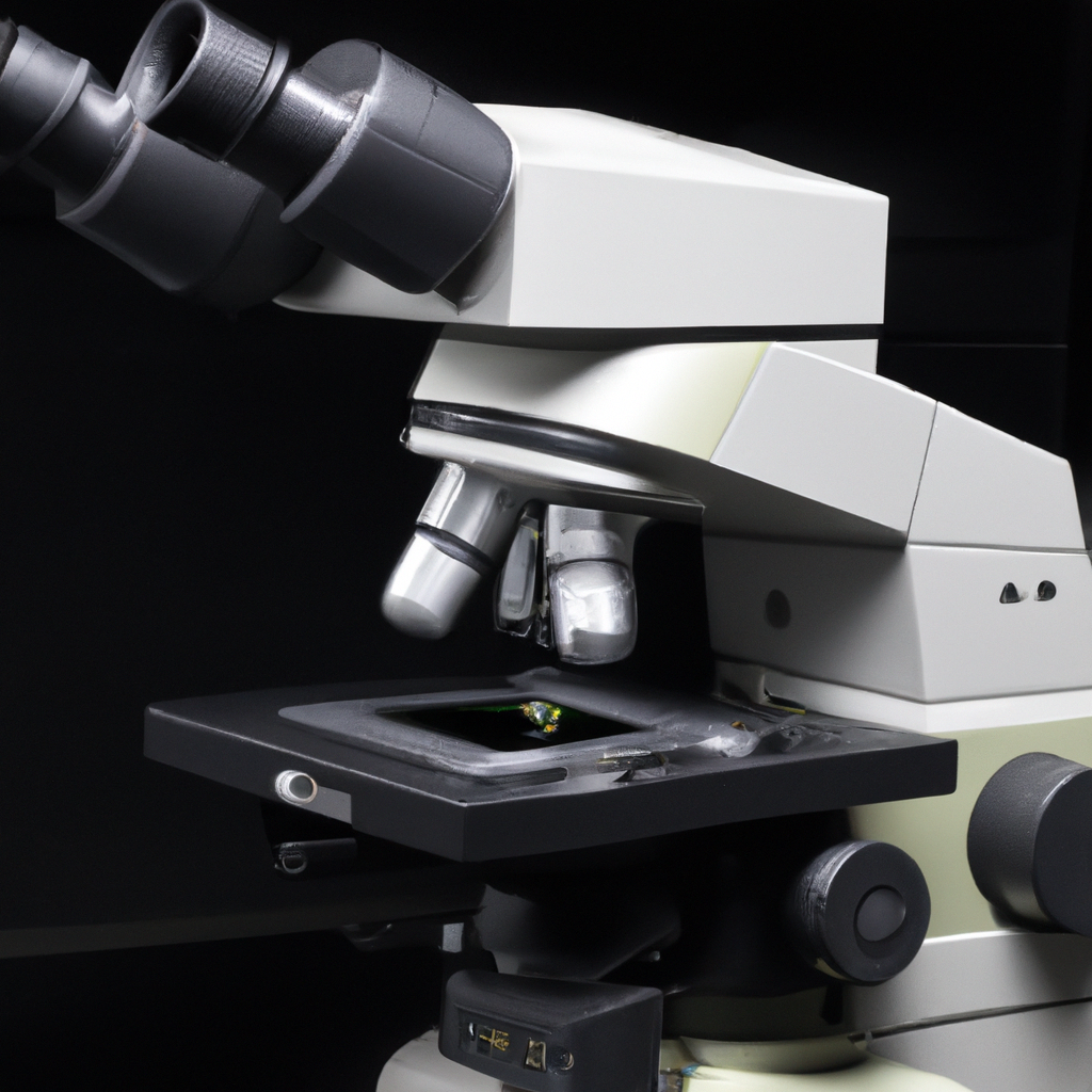
Are you curious about the benefits and drawbacks of a light optical microscope? This article will give you a brief overview of the advantages and disadvantages of using this type of microscope. Whether you are a student studying biology or a scientist conducting research, understanding the pros and cons of a light optical microscope can help you make informed decisions during your scientific explorations. So, let’s get started and explore the fascinating world of light optical microscopes!

Advantages of a light optical microscope
High magnification
One of the major advantages of a light optical microscope is its ability to provide high magnification. With proper lenses and adjustments, a light optical microscope can magnify the specimen up to 1000 times or more, allowing the observer to study the finest details of the sample. This high magnification is particularly useful for examining small structures like cells and their organelles, as well as for conducting detailed morphological analysis.
Easy to use
Another significant advantage of a light optical microscope is its user-friendly nature. Unlike more advanced microscopy techniques that require extensive training and technical knowledge, light optical microscopes are relatively easy to operate. They typically have simple controls and straightforward procedures for sample preparation and focusing. This makes them accessible to a wide range of users, including students, researchers, and professionals in various fields.
Real-time observation
Light optical microscopes offer the advantage of real-time observation, enabling scientists to directly observe the behavior and dynamics of the specimen in a live setting. This is particularly vital for studying living organisms, as it allows researchers to observe biological processes, cellular interactions, and dynamic changes in real-time. Real-time observation also provides the opportunity to capture and record important events as they occur, facilitating accurate data collection and analysis.
Affordable
Compared to other microscopy techniques, light optical microscopes are relatively affordable, making them accessible to individuals and institutions with limited budgets. The basic design and technology of these microscopes have been refined over the years, resulting in cost-effective models that still deliver reliable performance. The affordability factor allows for wider adoption of light optical microscopy across various fields, including education, research, and industry.
Can view living organisms
One significant advantage of a light optical microscope is its ability to observe and study living organisms. With the right configuration and techniques, researchers can observe the movement, growth, and interaction of living organisms without causing harm or disrupting their natural state. This opens up a whole realm of possibilities for studying biology, microbiology, and other life sciences, providing a valuable tool for exploring the intricacies of living systems.
Can be used with a wide range of samples
Light optical microscopes are versatile in terms of the types of samples that can be observed. They can be used with a wide range of specimens, including solid materials, liquids, and even gases. From biological samples like cells and tissues to geological samples or industrial materials, the adaptability of light optical microscopes allows for comprehensive examination across different fields. This versatility enhances the applicability of these microscopes in a variety of research areas.
Non-destructive imaging
A significant advantage of light optical microscopy is that it allows for non-destructive imaging. Unlike certain imaging techniques that may require sample manipulation or staining, light optical microscopes can examine samples without altering their properties or causing damage. This non-destructive nature ensures that the specimen remains intact and can be further analyzed or studied using other techniques if desired. It also minimizes any potential artifacts or distortions that may arise from sample preparation.
Versatility in illumination techniques
Light optical microscopes offer a wealth of options when it comes to illumination techniques. Different illumination methods, such as brightfield, darkfield, phase contrast, and fluorescence, can be utilized depending on the specific requirements of the observation. This versatility allows researchers to enhance contrast, highlight specific structures, or detect fluorescent labels in their samples. The ability to tailor illumination techniques to the specimen greatly enhances the level of detail and information that can be obtained from the microscope.
Small and portable
Light optical microscopes are often designed to be compact and portable, allowing for ease of use and transportation. This portability makes them suitable for various applications, including field studies and on-site analysis. Whether in a laboratory, a classroom, or in the field, the small and portable nature of light optical microscopes offers flexibility and convenience, enabling researchers and educators to bring the microscope to the desired location, rather than having to move or transport the samples.
Available in different configurations
Light optical microscopes are available in a variety of configurations to suit different needs and applications. From basic models for educational purposes to advanced research-grade instruments, there are options available for various budgets and requirements. Some microscopes may offer additional features such as digital imaging capabilities, motorized stages, or specialized objectives for specific applications. The availability of different configurations ensures that users can choose a microscope that best fits their specific needs and goals.
Disadvantages of a light optical microscope
Limited resolution
One of the limitations of light optical microscopy is its limited resolution. Resolution refers to the ability of a microscope to distinguish two closely spaced objects as separate entities. Light optical microscopes are subjected to the diffraction limit, which sets a theoretical limit on the smallest details that can be resolved. This means that, in some cases, smaller structures or fine details may not be visible under a light optical microscope. For higher resolution imaging, more advanced techniques such as electron microscopy are required.
Lower magnification compared to other microscopy techniques
While light optical microscopes offer high magnification capabilities, they generally have lower magnification limits compared to other microscopy techniques such as electron microscopy. Electron microscopes can achieve significantly higher magnifications, allowing for examination of ultra-fine structures at the nanoscale. Therefore, if the sample requires extremely high magnification, a light optical microscope may not be the ideal choice and other microscopy techniques should be considered.
Restricted to transparent or thin specimens
Light optical microscopes have limitations when it comes to the type of specimens that can be observed. These microscopes primarily rely on transmitted light, which means that they are best suited for transparent or thin specimens. Samples that are opaque or have a high degree of light absorption may not produce clear images or may not be suitable for observation under a light optical microscope. In such cases, alternative microscopy techniques like confocal microscopy or scanning electron microscopy may be more appropriate.
Brightness and contrast limitations
Another disadvantage of light optical microscopy is the limitation in achieving optimal brightness and contrast in certain specimens. While illumination techniques can enhance contrast to some extent, certain samples may still present challenges in terms of achieving optimum visibility. Issues such as low light levels, light scattering, or uneven illumination can impact the image quality and hinder the ability to observe fine details. It is important to carefully consider the sample characteristics and adjust the microscope settings accordingly to optimize the brightness and contrast during imaging.
Limited depth of field
Depth of field refers to the vertical range within a sample that remains in focus at a particular magnification. Light optical microscopes have a limited depth of field, which means that only a small portion of a three-dimensional sample can be in focus at a time. This limitation becomes more prominent when imaging thicker specimens or complex structures. If a sample has a large height or depth, it may be challenging to observe the entire structure in focus under a light optical microscope. This can restrict the level of detail that can be observed in certain samples.
Subject to diffraction limit
As mentioned earlier, light optical microscopes are subject to the diffraction limit, which is a fundamental limitation in imaging small structures or details. The diffraction of light imposes restrictions on the resolution that can be achieved, resulting in a limited ability to distinguish closely spaced objects. This diffraction limit often depends on the wavelength of the light used and the numerical aperture of the objective lens. While newer techniques and technologies have helped overcome this limitation to some extent, light optical microscopes still have inherent diffraction-based restrictions on their imaging capabilities.
Susceptible to optical distortions
Optical distortions can occur in light optical microscopes, impacting the quality and accuracy of the observed images. Various factors, such as lens aberrations, spherical aberrations, or chromatic aberrations, can introduce distortions in the image. These distortions can lead to blurred or distorted features, reducing the clarity and reliability of the observed data. Steps can be taken to minimize these distortions, such as using high-quality lenses and calibration, but it is essential to be aware that optical distortions can exist in light optical microscopy.
Staining and sample preparation required
In many cases, staining and sample preparation are necessary to optimize the visibility and contrast of the specimen under a light optical microscope. Staining techniques involve treating the sample with specific dyes or chemicals to enhance certain structures or highlight specific components. While staining can provide valuable information and improve the imaging quality, it also introduces additional steps and potential artifacts. Sample preparation itself can be time-consuming and may require specific protocols, depending on the nature of the specimen. These additional requirements may add complexity to the imaging process and increase the overall time and effort required.
Limited imaging capabilities in certain applications
Light optical microscopes may have limited imaging capabilities in specific applications or research areas. For example, when studying subcellular structures or molecular interactions at extremely small scales, the resolution and magnification limits of light optical microscopes may hinder detailed observation. Certain specialized techniques, such as super-resolution microscopy or electron microscopy, provide enhanced imaging capabilities to overcome these limitations. It is important to consider the specific requirements of the research or application and select the appropriate microscopy technique accordingly.
Limited use for subcellular structures
While light optical microscopes are invaluable tools for studying cell-level structures and behaviors, they have limitations when it comes to observing subcellular structures in detail. Subcellular structures such as mitochondria, endoplasmic reticulum, or individual proteins are often beyond the resolution capabilities of light optical microscopy. To visualize and study these ultra-small structures, techniques like electron microscopy or super-resolution microscopy are typically required. Therefore, light optical microscopes may not be the most suitable choice when analyzing the intricate components of subcellular biology.
In conclusion, light optical microscopes offer numerous advantages including high magnification, ease of use, real-time observation, affordability, and the ability to view living organisms. They are versatile in terms of the range of samples they can be used with and provide non-destructive imaging. Additionally, light optical microscopes offer versatility in illumination techniques, are small and portable, and available in different configurations to suit specific needs. However, light optical microscopes also have their limitations, including limited resolution, lower magnification compared to other microscopy techniques, restriction to transparent or thin specimens, brightness and contrast limitations, limited depth of field, subject to the diffraction limit, susceptibility to optical distortions, staining and sample preparation requirements, limited imaging capabilities in certain applications, and limited use for subcellular structures. Understanding these advantages and disadvantages helps researchers and users make informed choices when selecting a microscopy technique for their specific needs.


