Have you ever wondered about the hidden wonders of the microscopic world? Curiosity arises when we are faced with the incredible diversity and complexity of organisms, cells, and molecules that make up our natural surroundings. It is this curiosity that has fueled the advent of light microscopes, allowing us to explore the smallest of details within the living and non-living world. By harnessing the power of light, these remarkable instruments have revolutionized scientific research and discovery, providing us with a multitude of benefits that continue to shape our understanding of the world around us.

Enhanced visibility
Clear and sharp images
Using a light microscope offers enhanced visibility through clear and sharp images. The optical system of a light microscope enables the user to observe specimens with exceptional clarity. The lenses and illuminating system work together to produce well-defined images, allowing for detailed examination of objects under observation. Whether you are studying cells, tissues, or small organisms, the clear and sharp images produced by a light microscope facilitate accurate analysis and interpretation.
Ability to observe living specimens
One of the great benefits of a light microscope is its ability to observe living specimens. Unlike electron microscopes, which require samples to be fixed and prepared, light microscopes allow for real-time observation of organisms and cells. This is particularly valuable in fields such as biology and medicine, where the study of living organisms is essential. By being able to observe organisms in their natural state, researchers and scientists can gain a deeper understanding of biological processes and behaviors.
Visualizing cellular structures and organelles
Light microscopes enable the visualization of cellular structures and organelles, which are integral to the functioning of living organisms. With the aid of staining techniques and suitable magnification levels, researchers can examine the intricate details of cells and their components. This allows for the study of cell division, organelle function, and other cellular processes. By visualizing and understanding cellular structures, scientists can make significant contributions to fields such as cell biology, genetics, and developmental biology.
Distinguishing different staining techniques
Another advantage of a light microscope is its ability to distinguish different staining techniques. Staining is a common practice used to enhance the contrast and visibility of cellular structures and tissues. With a light microscope, researchers can distinguish between different staining methods, enabling them to accurately interpret the results of their experiments. This capability is particularly important in fields such as histology and pathology, where the identification and classification of tissues rely heavily on staining techniques.
Convenience and cost-effectiveness
Portability and ease of use
Light microscopes are known for their portability and ease of use. Unlike larger and more complex imaging equipment, light microscopes can be easily transported from one location to another. This portability makes them particularly suitable for fieldwork, where researchers may need to analyze specimens on-site. Additionally, light microscopes are relatively straightforward to operate, making them accessible to individuals with varying levels of microscopy experience. The ease of use and portability of light microscopes enhance their convenience and practicality in various scientific settings.
Affordable and accessible equipment
Compared to other microscopy techniques, light microscopes are generally more affordable and accessible. Their simple design and use of basic optical principles allow for cost-effective manufacturing, making them a viable option for educational institutions, research laboratories, and even hobbyists. Furthermore, the widespread use and popularity of light microscopes mean that there is a wide range of models and brands available on the market, catering to different budgets and requirements. This accessibility ensures that individuals and organizations can readily obtain a light microscope without breaking the bank.
Minimal maintenance and operational costs
Maintaining and operating a light microscope typically incurs minimal costs. Unlike more advanced imaging techniques, light microscopes do not require expensive consumables or regular servicing. The basic maintenance tasks, such as cleaning the lenses and adjusting the alignment, can usually be done by the user without the need for professional assistance. Additionally, the operational costs of a light microscope, such as power consumption and bulb replacement, are generally low. These factors contribute to the overall cost-effectiveness of using a light microscope for scientific research and educational purposes.
Low power requirements
Another cost-saving aspect of light microscopes is their low power requirements. Unlike electron microscopes that require high-energy electron beams, light microscopes operate using simple light sources, typically an LED or halogen bulb. This low power consumption not only results in lower electricity bills but also reduces the environmental impact. Light microscopes can be used in various settings, including fieldwork in remote areas or resource-limited regions where access to stable power supply might be a challenge. The low power requirements of light microscopes ensure their versatility and practicality in diverse research and educational contexts.
Versatility and flexibility
Wide range of magnification levels
Light microscopes offer a wide range of magnification levels, allowing for the examination of objects at different scales. Depending on the specific objectives and lenses used, users can achieve magnifications from as low as 40x to as high as 1000x or more. This range of magnification provides the flexibility to observe a variety of specimens, from large macroscopic structures to tiny microorganisms. Researchers and educators can tailor the magnification levels to suit their specific study objectives, enabling them to explore the microscopic world in extensive detail.
Ability to switch between different objectives
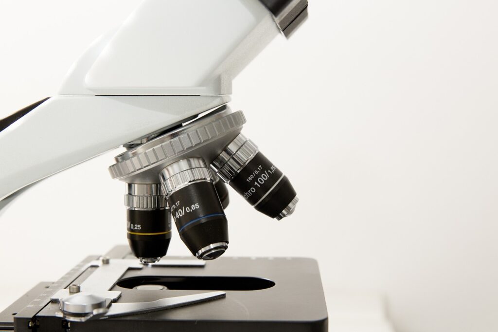
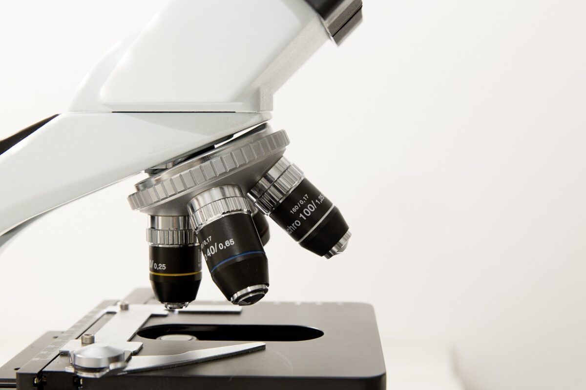
Another advantage of light microscopes is the ability to switch between different objectives. Objectives are the lenses that determine the magnification and resolution of the microscope. Light microscopes are typically equipped with multiple objectives, each with a different magnification level. By simply rotating the nosepiece, users can easily switch between objectives, enabling a seamless transition between different levels of magnification. This versatility is beneficial when studying specimens with varying sizes or when needing to focus on different aspects of the sample during observation.
Observing various sample types
Light microscopes are compatible with various sample types, making them highly versatile instruments. Whether you are examining a thin section of tissue on a glass slide, a prepared wet mount of living organisms, or a stained smear, a light microscope can accommodate different sample types. This versatility allows researchers and educators to study a wide range of biological materials, including plant tissues, animal cells, blood samples, and microorganisms. Being able to observe various sample types is essential in many scientific disciplines, allowing for comprehensive research and analysis.
Compatibility with different specimen preparation techniques
Light microscopes are compatible with different specimen preparation techniques, offering flexibility in experimental approaches. From simple wet mounts to complex staining methods, researchers have multiple options for preparing their samples for microscopy. This compatibility facilitates the integration of various techniques, such as phase contrast, fluorescence, and differential interference contrast (DIC), into the observation process. By being able to choose the most appropriate specimen preparation technique, researchers can optimize the visualization of specific structures or enhance the contrast of certain components, ultimately leading to more insightful observations and discoveries.
Real-time observation
Time-lapse imaging of dynamic processes
One of the most valuable features of light microscopes is their capability for time-lapse imaging of dynamic processes. Time-lapse microscopy involves capturing a series of images over a designated period, allowing for the visualization of changes and movements that occur over time. This technique is especially useful for studying cellular processes, such as cell division, migration, and morphological changes. By capturing real-time images at regular intervals, researchers can analyze the dynamics and kinetics of biological events, providing a deeper understanding of their underlying mechanisms.
Continuous monitoring of cell behavior
Light microscopes enable continuous monitoring of cell behavior, providing valuable insights into cellular activities. By observing cells in real time, researchers can track and analyze their responses to stimuli, environmental changes, or therapeutic interventions. This continuous monitoring enhances our understanding of cell signaling pathways, cell kinetics, and cell-cell interactions, among other biological phenomena. Moreover, the ability to continuously monitor cell behavior allows researchers to capture rare or transient events that might be missed in static observations, leading to novel discoveries and scientific breakthroughs.
Following changes over extended periods
Light microscopes also allow for the following of changes over extended periods, providing a long-term perspective on biological phenomena. By repeatedly observing the same sample over multiple days or weeks, researchers can track the progression of events, such as tissue growth, cellular differentiation, or the effects of aging. This longitudinal approach aids in uncovering patterns, trends, and developmental processes that would not be apparent through a single time point observation. Following changes over extended periods adds a valuable temporal dimension to scientific research and helps elucidate complex biological processes.
Real-time interaction and adjustments
In addition to capturing and analyzing images, light microscopes enable real-time interaction and adjustments during observation. This feature is particularly useful when researchers need to make immediate changes to focus, lighting, or other microscope settings. By being able to interact with the live image on the microscope’s monitor, scientists can optimize the quality of the observation, ensuring that the most relevant information is captured. Real-time interaction also facilitates collaboration and sharing of findings during live microscopy sessions, promoting interdisciplinary discussion and knowledge exchange.

Non-destructive analysis
Preserving integrity of samples
Light microscopy techniques allow for non-destructive analysis, preserving the integrity of samples. Unlike some other analytical techniques that require destructive sample preparation or irreversible changes, light microscopes can observe specimens without causing significant damage. This non-destructive nature is particularly crucial when working with fragile or valuable samples, such as rare specimens or irreplaceable archival materials. Preserving the integrity of samples allows for further analysis or potential future use in other research endeavors, ensuring the long-term availability of valuable scientific resources.
Reducing the need for excessive sample preparation
Using a light microscope reduces the need for excessive sample preparation, saving time and resources. While certain sample preparation techniques may still be required, light microscopes can often provide meaningful observations with minimal intervention. Compared to techniques like electron microscopy, which typically involve intricate sample preparation processes, light microscopy allows for more immediate analysis. This streamlined workflow reduces the time and effort invested in preparing samples, enabling researchers to focus on the actual observation and interpretation of the results.
Non-invasive visualization of tissues and cells
Light microscopy offers non-invasive visualization of tissues and cells, avoiding interference with their natural state. Because light microscopes utilize non-ionizing radiation (light) to illuminate specimens, they do not harm biological materials during the observation process. This non-invasive nature is crucial for studying living organisms, as it allows researchers to explore their biology without adversely affecting their viability or functionality. Non-invasive visualization is particularly relevant in areas such as developmental biology, where the observation of live embryos, tissues, and organs is essential for understanding growth and morphological changes.
Minimizing damage to delicate or valuable specimens
Light microscopy minimizes damage to delicate or valuable specimens, ensuring their long-term integrity. When working with sensitive samples, such as fragile tissues, rare specimens, or artifacts, it is crucial to minimize any potential harm or degradation. Light microscopes, through their gentle imaging conditions and non-invasive nature, help protect such specimens from unnecessary damage or deterioration. By utilizing appropriate imaging techniques and careful handling, researchers can maintain the quality and longevity of delicate or valuable materials, preserving them for future scientific inquiries or historical preservation.
Educational value
Introduction to basic biological concepts
Light microscopy holds significant educational value by introducing students to basic biological concepts. Through the observation of various biological specimens, students can develop an understanding of cellular structures, tissue organization, and organismal complexity. By visualizing living organisms and cellular processes, students can grasp fundamental principles such as mitosis, photosynthesis, or the structure-function relationships of different cell types. Light microscopes provide a tangible and engaging way to learn about biology, fostering curiosity, and laying the foundation for deeper scientific exploration.
Teaching fundamental microscopy skills
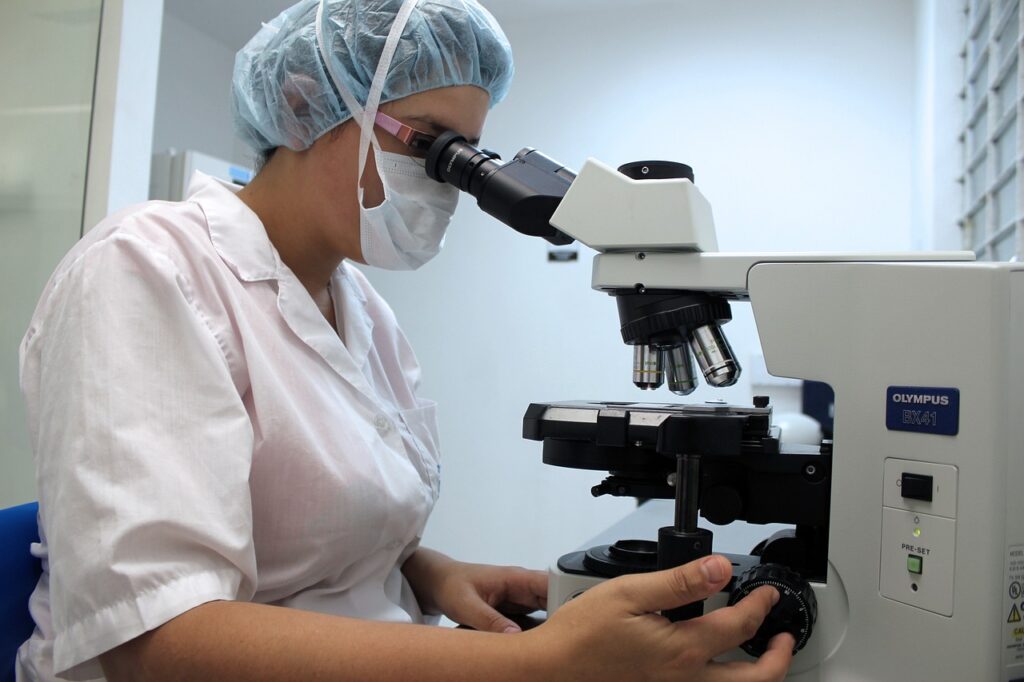
Using a light microscope provides an excellent opportunity to teach students fundamental microscopy skills. Learning how to correctly operate a microscope, adjust the focus, and navigate through different magnifications are essential laboratory techniques. By gaining proficiency in microscopy, students acquire valuable transferable skills applicable across various scientific disciplines. Microscopy skills go beyond biology and can be relevant in fields such as materials science, forensic analysis, or environmental monitoring. Teaching these skills through light microscopy promotes scientific literacy, critical thinking, and observational abilities in students.
Engaging students through hands-on exploration
Light microscopes engage students through hands-on exploration, fostering their passion for science. The ability to observe living organisms, view intricate cellular structures, and make scientific discoveries firsthand captivates students’ curiosity. Actively using a light microscope empowers students to take an active role in their learning, promoting a deeper understanding and appreciation for the natural world. The hands-on nature of light microscopy also encourages collaboration, experimentation, and problem-solving skills, all of which are crucial foundations for future scientific endeavors.
Illustrating scientific principles and discoveries
Light microscopes serve as powerful tools for illustrating scientific principles and discoveries. By directly observing the microscopic world, students can witness the mechanisms behind scientific phenomena and theories. From observing cell division to visualizing photosynthetic processes, light microscopes provide concrete evidence of scientific principles. Additionally, light microscopy can showcase historical scientific discoveries, such as the observation of bacteria by Antonie van Leeuwenhoek or the identification of DNA’s double helix structure by James Watson and Francis Crick. The visual impact of light microscopy aids in conveying scientific concepts and fostering a sense of wonder and appreciation for discovery.

Applications in research
Investigation of disease pathology
Light microscopy plays a critical role in the investigation of disease pathology. By examining tissues and cells under a light microscope, researchers can identify pathological changes associated with various diseases. Histological analysis of biopsy samples can reveal abnormalities, such as cellular dysplasia or the presence of cancerous cells. In combination with staining techniques, light microscopes aid in the detection and classification of diseases, contributing to accurate diagnosis and treatment planning. The ability to study disease pathology at the cellular level enhances our understanding of diseases and paves the way for the development of targeted therapeutics.
Study of cellular and molecular processes
The study of cellular and molecular processes heavily relies on light microscopy. Light microscopes enable researchers to investigate a wide range of cellular activities, such as protein localization, signaling pathways, and gene expression. By using fluorescent probes and markers, researchers can label specific molecules or structures within cells, allowing for detailed visualization and analysis. This enables the examination of intracellular dynamics, interactions between molecules, and the effects of genetic or environmental factors on cellular processes. Light microscopy provides a valuable tool for unraveling the complexities of cellular and molecular biology.
Exploration of genetic and developmental mechanisms
Understanding genetic and developmental mechanisms is crucial in biology, and light microscopy facilitates this exploration. By studying model organisms, such as fruit flies or zebrafish embryos, researchers can observe various stages of development under a light microscope. This allows for the identification of genetic factors that influence embryonic development, morphogenesis, and organogenesis. The ability to visualize and dissect these processes contributes to our understanding of developmental biology and provides insights into potential interventions for developmental disorders or birth defects.
Evaluation of drug effects on living organisms
Light microscopy plays a significant role in evaluating drug effects on living organisms. Researchers can use light microscopes to observe the response of cells or organisms to new pharmaceutical compounds or therapeutic interventions. Through time-lapse imaging or repeated observations, researchers can track changes in cell behavior, tissue regeneration, or the effects of drugs on disease progression. Light microscopy allows for in vivo or ex vivo evaluation of drug efficacy, side effects, or potential toxicities. With this information, researchers can make informed decisions regarding drug development and optimize treatment strategies.
Interdisciplinary utility
Use in medical, biological, and environmental fields
Light microscopy finds utility across various scientific disciplines, including medicine, biology, and environmental studies. In the medical field, light microscopes aid in diagnosing diseases, analyzing tissue samples, and monitoring treatment responses. In biology, light microscopy is indispensable for studying cellular processes, ecological interactions, or biodiversity. Environmental scientists use light microscopes to analyze soil samples, study microorganisms in aquatic ecosystems, or monitor pollution levels. The interdisciplinary utility of light microscopy highlights its versatility and widespread applicability, making it an essential tool in numerous scientific endeavors.
Integration with other analytical techniques
Light microscopy can be seamlessly integrated with other analytical techniques, enhancing scientific inquiry. For example, researchers can combine light microscopy with immunohistochemistry or molecular biology techniques to investigate the expression patterns of specific proteins or genes. Furthermore, the integration of light microscopy with advanced imaging modalities, such as confocal microscopy or super-resolution microscopy, allows for higher resolution and improved 3D visualization of biological structures. By integrating different techniques, researchers can obtain a comprehensive understanding of complex biological processes or obtain complementary information that would be difficult to access using a single technique.
Complementing other imaging modalities
In addition to integrating with analytical techniques, light microscopy can complement other imaging modalities. For instance, while electron microscopy provides unparalleled resolution and allows for detailed examination of ultrastructures, it lacks the ability to observe living specimens. Light microscopy fills this gap by enabling non-invasive imaging of dynamic processes in real-time. By combining the strengths of different imaging modalities, researchers can obtain a more comprehensive view of biological phenomena, unraveling multiple layers of information and enriching scientific discoveries.
Contributing to multiple scientific disciplines
Light microscopy makes significant contributions to multiple scientific disciplines and fosters collaboration between researchers from different fields. Scientists from diverse backgrounds, such as medicine, physics, chemistry, or ecology, can all benefit from utilizing light microscopes in their research. By facilitating communication and knowledge exchange, light microscopy drives interdisciplinary research and promotes a holistic understanding of complex scientific problems. The application of light microscopy across various scientific disciplines sparks innovation, generates novel insights, and ultimately advances our collective knowledge and understanding of the natural world.
Training and skill development
Learning essential laboratory techniques
Using a light microscope provides an opportunity to learn essential laboratory techniques. Proper handling of microscope slides, sample preparation, and maintenance of microscope equipment are crucial skills for anyone working in a laboratory setting. Learning these techniques through light microscopy builds a solid foundation for future scientific endeavors and fosters good laboratory practices. Understanding laboratory techniques is not only valuable for research or educational purposes but also for professional development and career advancement in scientific fields.
Gaining proficiency in microscopy
Proficiency in microscopy is a valuable skill that can be gained through using light microscopes. By regularly practicing observation, adjusting focus, and navigating through different magnifications, individuals can become skilled microscope users. Developing proficiency in microscopy enables researchers and educators to confidently utilize this tool for their scientific inquiries or teaching activities. Proficiency in microscopy goes beyond technical know-how and contributes to critical thinking, problem-solving, and attention to detail, essential qualities for successful scientific endeavors.
Developing attention to detail and patience
Working with light microscopes cultivates attention to detail and patience, key attributes for scientific research. The observation of microscopic details requires careful scrutiny and an ability to detect subtle changes or variations. Researchers using light microscopes develop a keen eye for detail, enabling them to notice important features that might otherwise go unnoticed. Furthermore, light microscopy often involves time-consuming processes, such as sample preparation or capturing time-lapse images over extended periods. This requires patience and dedication, as scientific discoveries often come through meticulous and methodical observation.
Understanding image analysis and interpretation
Using light microscopes enhances understanding of image analysis and interpretation. Light microscopy generates a vast amount of image data, and researchers must possess the skills to effectively analyze and interpret this information. This includes distinguishing cells or structures of interest, quantifying variables, and drawing meaningful conclusions from the observed data. By practicing image analysis and interpretation, individuals develop a critical mindset and an ability to draw informed conclusions from complex datasets. Understanding image analysis is essential not only in microscopy but also in various scientific disciplines that rely on visual data interpretation.
Historical significance
Foundation of modern microscopy
Light microscopy holds historical significance as the foundation of modern microscopy. The invention of the light microscope in the 17th century by pioneers like Antonie van Leeuwenhoek laid the groundwork for scientific exploration at the microscopic level. These early microscopes utilized simple lenses and light sources, opening up the microscopic world for observation. Over the centuries, advancements in optics and technologies have led to the development of more sophisticated light microscopes, enabling higher resolution and improved image quality. The historical significance of light microscopy cannot be overstated, as it paved the way for subsequent advancements in microscopy techniques.
Contributions to scientific discoveries
Light microscopy has made significant contributions to numerous scientific discoveries and breakthroughs. From the early observations of microorganisms by Antonie van Leeuwenhoek to the identification of chromosomes and DNA, light microscopy has played a pivotal role in expanding our understanding of the natural world. Countless anatomical and physiological discoveries have been made using light microscopes, providing insights into biological mechanisms and shaping our knowledge of life on Earth. The contributions of light microscopy to scientific discoveries demonstrate its enduring impact on scientific progress and our understanding of the world around us.
Pioneering advancements in optics
The development of light microscopy has driven pioneering advancements in optics. Researchers and inventors have continually pushed the boundaries of optical design, resulting in improved resolution, contrast, and imaging capabilities. From the introduction of achromatic lenses to the development of phase contrast microscopy and fluorescence microscopy, each advancement in optical technology has expanded the possibilities of light microscopy. The pursuit of better optics has not only increased the power and versatility of light microscopes but has also influenced other fields, such as astronomy, spectroscopy, and telecommunications.
Evolution of microscopy techniques
Light microscopy has continuously evolved and diversified, giving rise to various microscopy techniques. Over time, different microscopy methods and modalities have emerged to address specific research needs or overcome limitations. Techniques such as confocal microscopy, multiphoton microscopy, and super-resolution microscopy have revolutionized the field of light microscopy. These advancements have pushed the boundaries of optical resolution, enabled 3D imaging, and allowed for the visualization of nanoscale structures. The evolution of microscopy techniques illustrates the dynamic nature of scientific progress, where new innovations build upon existing foundations and drive continuous improvement.


