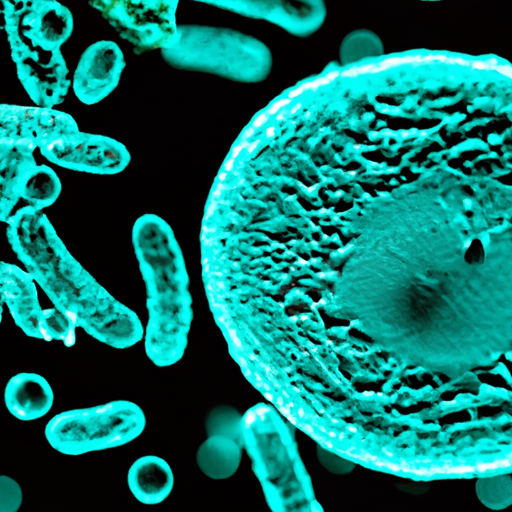
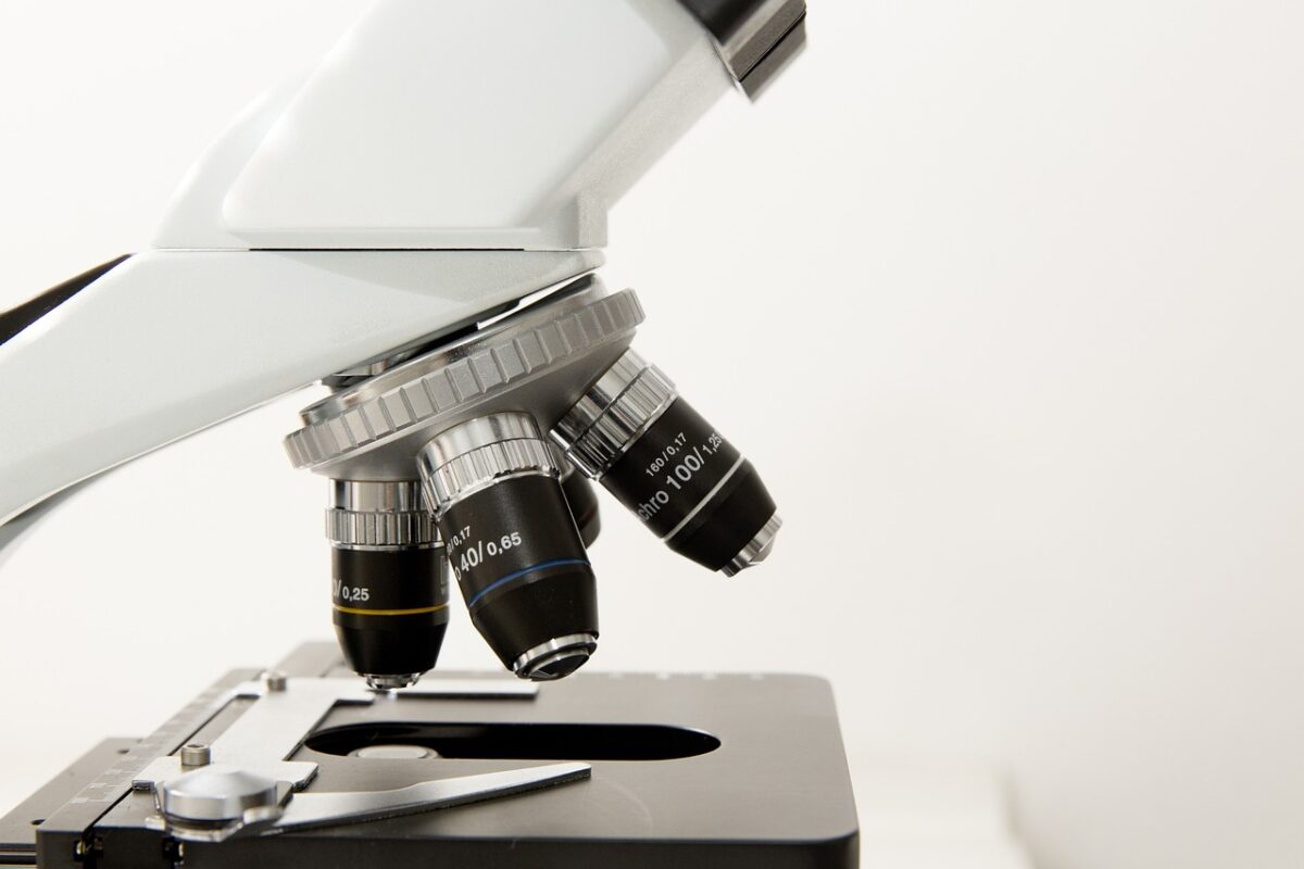
Imagine being able to explore the microscopic world, unraveling its mysteries right from the comfort of your home or laboratory. With the advent of digital microscopes, this once-difficult task has become a breeze. These innovative devices grant you the power to visualize bacteria with astonishing clarity, offering a new level of understanding and fascination. Gone are the days of squinting through traditional microscope eyepieces, as digital microscopes bring bacteria to life on your computer screen. This article will delve into the remarkable capabilities of digital microscopes and how they can revolutionize your exploration and study of bacteria.
Benefits of Using a Digital Microscope
Enhanced Visualization:
One of the major benefits of using a digital microscope is the enhanced visualization it provides. With a traditional microscope, you have to look through the eyepiece to observe the sample. However, with a digital microscope, you can view the sample on a high-resolution digital display. This enables you to see the bacteria with greater clarity, making it easier to identify and study their characteristics.
Image Capture and Sharing:
Another advantage of using a digital microscope is the ability to capture and share images of the bacteria. Digital microscopes are equipped with cameras that allow you to capture still images and even record videos of the bacteria in real-time. This is particularly useful for documentation purposes or for sharing your findings with colleagues or experts for further analysis.
Real-time Viewing:
Digital microscopes provide real-time viewing, which means you can observe the bacteria as they move and interact with their environment. This is especially beneficial when studying live bacteria or when trying to understand their behavior and response to different conditions. Real-time viewing allows for more accurate and dynamic observation, providing valuable insights into bacterial activities.
Measurement and Analysis:
Digital microscopes often come with measurement and analysis tools that enable you to quantify various characteristics of the bacteria. These tools allow you to measure the size, shape, and density of the bacteria, providing valuable data for further research. Additionally, digital microscopes can also perform image analysis, such as counting the number of bacteria in a specific area, which can facilitate quantitative analysis and comparison.
Ease of Use:
Digital microscopes are designed to be user-friendly, making them accessible to researchers and professionals of all levels of expertise. They often have intuitive interfaces and controls, allowing for easy adjustment of settings like magnification, focus, and contrast. This ease of use not only improves efficiency but also reduces the learning curve associated with traditional microscopy techniques.
Digital Microscope Technology
How Digital Microscopes Work:
Digital microscopes use a combination of lenses, sensors, and image processing technology to produce high-resolution images of the bacteria. The lenses in a digital microscope focus the light passing through the sample onto an image sensor, which captures the image and converts it into digital data. This data is then processed and displayed on a monitor, providing a clear and detailed view of the bacteria.
Advancements in Digital Microscopy:
Digital microscopy technology has advanced significantly over the years, leading to improvements in image quality and functionality. One such advancement is the introduction of high-resolution image sensors, which enable the capture of more detailed images. Additionally, advancements in image processing algorithms have made it possible to enhance and manipulate the captured images, further improving the visualization and analysis capabilities of digital microscopes.
Different Types of Digital Microscopes:
There are various types of digital microscopes available, each catering to different research needs and applications. Some common types include:
-
Compound Digital Microscopes: These microscopes are similar to traditional compound microscopes but are equipped with a digital camera for image capture and display.
-
Stereo Digital Microscopes: Stereo digital microscopes provide a three-dimensional view of the bacteria, allowing for better depth perception and spatial understanding.
-
USB Digital Microscopes: USB digital microscopes are portable and connect directly to a computer via a USB port, allowing for easy image capture and analysis.
-
Fluorescence Digital Microscopes: Fluorescence digital microscopes use fluorescent dyes to label bacteria, making specific structures or proteins visible under specific wavelengths of light.
Preparing Samples for Digital Microscopy
Collection and Culturing of Bacteria:
Before using a digital microscope, it is important to collect and culture the bacteria you wish to visualize. This typically involves obtaining a sample from the desired source, such as a swab from a surface or a liquid sample. The collected sample is then cultured in a suitable growth medium to encourage bacterial growth and multiplication. Culturing the bacteria ensures that you have a sufficient number of cells to visualize and study under the microscope.
Fixation and Staining Techniques:
To prepare bacteria samples for digital microscopy, fixation and staining techniques are often employed. Fixation involves treating the bacteria with chemicals to preserve their structure and prevent decay. Staining, on the other hand, involves the application of specific dyes or stains that selectively bind to different cellular components, making them more visible under the microscope. These techniques enhance the contrast and visibility of the bacteria, enabling more detailed observation and analysis.
Features and Functions of Digital Microscopes
Resolution and Magnification:
The resolution and magnification capabilities of a digital microscope are crucial for obtaining clear and detailed images of the bacteria. Resolution refers to the level of detail that can be captured, while magnification determines the apparent size of the bacteria. Higher resolution and magnification allow for more precise observation and analysis, enabling researchers to study even the smallest details of bacteria morphology and behavior.
Illumination and Contrast:
Digital microscopes employ various methods of illumination to enhance the visibility of the bacteria. Brightfield illumination, where the sample is illuminated from below, is commonly used for general observation. Additionally, advanced techniques such as darkfield and phase contrast illumination are used to improve contrast and highlight specific features of the bacteria that may be difficult to see under brightfield illumination.
Focusing Options:
Digital microscopes offer different focusing options to ensure clear and sharp images. These options may include manual focusing, where the user adjusts the focus manually, or autofocus, which automatically adjusts the focus based on the sample. Some digital microscopes also provide extended depth of field (EDF) imaging, where multiple images taken at different focal planes are combined to create one fully focused image.
Image Sensor and Processing:
The image sensor plays a crucial role in digital microscopy as it is responsible for capturing the image of the bacteria. Higher-quality image sensors can capture more detailed and accurate images, resulting in better visualization and analysis. Additionally, the image data captured by the sensor is processed by the microscope’s internal software, which may include various image enhancement algorithms to improve the visibility and quality of the captured images.
Software and Connectivity:
Digital microscopes are often equipped with software that allows for image capture, measurement, analysis, and even advanced functions like stitching together multiple images to create a panoramic view. This software is typically user-friendly, with intuitive interfaces and options for customization. Furthermore, digital microscopes may offer connectivity options such as USB or Wi-Fi, enabling easy transfer of data to other devices or networks.
Choosing the Right Digital Microscope for Bacteria Visualization
Application Requirements:
When choosing a digital microscope for bacteria visualization, it is crucial to consider your specific application requirements. Different research fields or applications may have different needs in terms of resolution, magnification, and imaging techniques. For example, if you are studying bacteria at the cellular level, you may require a microscope with higher magnification and resolution capabilities.
Budget and Cost Considerations:
Budget considerations are also an important factor when selecting a digital microscope. Digital microscopes vary in price depending on their features, capabilities, and brand. It is essential to evaluate your budget and opt for a microscope that provides the necessary features within your financial limitations. Additionally, consider long-term costs, such as maintenance and software updates, to ensure cost-effectiveness in the long run.
Ergonomics and Portability:
Ergonomics and portability should not be overlooked when choosing a digital microscope. It is important to select a microscope that offers comfort during prolonged use, as well as adjustable features to accommodate different users. Portability may be a crucial factor if you need to move or transport the microscope to different locations. Take into consideration the weight, size, and ease of setup and use when assessing the microscope’s portability.
Integration with Existing Equipment:
If you already have existing laboratory equipment, such as imaging or analysis systems, it is important to ensure compatibility and integration with your digital microscope. Integration with existing equipment can streamline workflows and data analysis, saving time and effort. Check if the microscope software allows for seamless data transfer or if there are available connectors or adapters to connect the microscope with your existing devices.
Techniques for Visualizing Bacteria Using a Digital Microscope
Direct Observation of Live Bacteria:
One technique for visualizing bacteria using a digital microscope is through direct observation of live bacteria. This involves placing a small sample containing live bacteria onto a microscope slide or petri dish and directly viewing them under the microscope’s high-resolution camera. This technique enables you to observe the bacteria in their natural state, seeing how they move, reproduce, and interact with their environment.
Visualization of Stained Bacteria:
Staining is a widely used technique to enhance the visibility of bacteria under a digital microscope. Different stains or dyes can be applied to the bacteria, selectively binding to different cellular components and making them more visible. Common staining techniques include Gram staining, which categorizes bacteria into Gram-positive and Gram-negative, and fluorescent staining, which uses fluorescent dyes to label specific structures or proteins within the bacteria.
Phase Contrast and Differential Interference Contrast Imaging:
Phase contrast and differential interference contrast (DIC) imaging are advanced techniques that enhance the contrast and visibility of bacteria without the need for staining. These techniques take advantage of the differences in refractive index between the bacteria and their surroundings, resulting in a clearer image. Phase contrast and DIC imaging are particularly useful when studying live bacteria or delicate samples that may be damaged by staining techniques.

Image Processing and Analysis
Image Enhancement:
Image enhancement techniques can be applied to improve the quality and visibility of the captured images. These techniques include adjusting brightness, contrast, and color balance, as well as reducing image noise and artifacts. Image enhancement allows for better visualization and analysis of the bacteria, especially when dealing with low-contrast or noisy images.
Measurement and Quantification:
Digital microscopes often come with measurement and quantification tools that allow for the precise analysis of bacteria characteristics. These tools enable the measurement of various parameters, such as size, shape, and density of bacteria. Quantitative analysis provides valuable data for statistical comparison, trend analysis, and further research.
Automated Detection and Classification:
Advanced digital microscopes may offer automated detection and classification capabilities, using image analysis algorithms to identify and classify bacteria automatically. These algorithms can detect and count bacteria, classify them based on certain criteria, or even identify specific strains or species. Automated detection and classification save time and effort compared to manual analysis, especially when dealing with a large number of bacteria samples.
Challenges and Limitations of Digital Microscopy for Bacteria
Resolution Constraints:
While digital microscopes have significantly improved resolution compared to traditional microscopes, there are still limitations when visualizing bacteria at extremely small scales. Some bacteria may have features or structures that are smaller than the resolution limit of the microscope, making them challenging to analyze. Overcoming resolution constraints often requires more advanced imaging techniques or higher magnification capabilities.
Sampling Artifacts:
Sample preparation for digital microscopy, including fixation and staining, can introduce artifacts that may affect the accuracy of bacterial visualization. Artifacts include shrinkage, distortion, or alterations in the bacteria’s morphology or structure. It is essential to be aware of these potential artifacts and minimize their impact through careful sample preparation techniques.
Limitations of Staining Techniques:
While staining is a widely used technique for enhancing bacterial visibility, certain bacteria do not stain well, resulting in poor contrast and visibility. Additionally, staining techniques may alter the properties or behavior of the bacteria, potentially affecting the accuracy of observations. It is important to select the appropriate staining technique and consider its limitations when visualizing specific bacteria species or strains.
Applications of Digital Microscopy in Bacteria Research
Microbial Ecology and Environmental Studies:
Digital microscopy has revolutionized the field of microbial ecology and environmental studies by enabling researchers to study bacteria in their natural habitats. It allows for the observation and characterization of bacteria in various environmental samples, such as soil, water, or air. Understanding the ecological role and behavior of bacteria is crucial for assessing ecosystem dynamics, nutrient cycling, and climate change impacts.
Medical and Clinical Diagnosis:
Digital microscopy plays a vital role in medical and clinical diagnosis, particularly in the identification and characterization of bacterial pathogens. It allows for the rapid and accurate detection of bacteria in clinical samples, aiding in the diagnosis of infectious diseases. Digital microscopy can also assist in monitoring treatment efficacy and studying bacterial resistance patterns, contributing to the development of effective treatment strategies.
Food and Beverage Quality Control:
The food and beverage industry relies on digital microscopy for quality control and safety assessments. Digital microscopes can be used to detect and identify bacteria in food and beverage products, ensuring compliance with safety standards and regulations. Microscopic examination can help identify spoilage bacteria, assess product contamination, and monitor microbial growth during food processing and storage.
Conclusion
The use of digital microscopes has revolutionized the field of bacteria visualization and analysis. These advanced instruments offer enhanced visualization capabilities, allowing for detailed observation and measurement of bacteria. With features such as image capture, real-time viewing, and image analysis, digital microscopes provide valuable tools for researchers in various fields, from microbial ecology to medical diagnosis. While there are challenges and limitations associated with digital microscopy, the benefits and applications far outweigh these concerns. Digital microscopes have become indispensable tools in the study of bacteria, enabling researchers to gain valuable insights into their structure, behavior, and ecological roles.
